Imaging

Dental Radiographs (X-rays)
Dental X-rays are valuable diagnostic tools that allow us to safely and effectively evaluate the health of the jaw bones, tooth roots, and contact areas where the teeth touch each other--all areas we cannot observe directly with our eyes. We search for hidden dental diseases such as cavities, gum and bone disease, abscesses, cysts, and tumors. X-rays are also used to view the progress of eruption of permanent teeth in children, for orthodontic and implant treatment planning, and for many other dental procedures.
Digital X-rays are safer, faster, more accurate, and more environmentally friendly than traditional film X-rays. With limited radiation exposure and instantaneous images that are clearer and more accurate, they improve diagnosis and patient care.
Digital X-rays are safer, faster, more accurate, and more environmentally friendly than traditional film X-rays. With limited radiation exposure and instantaneous images that are clearer and more accurate, they improve diagnosis and patient care.
Cone Beam Scanner
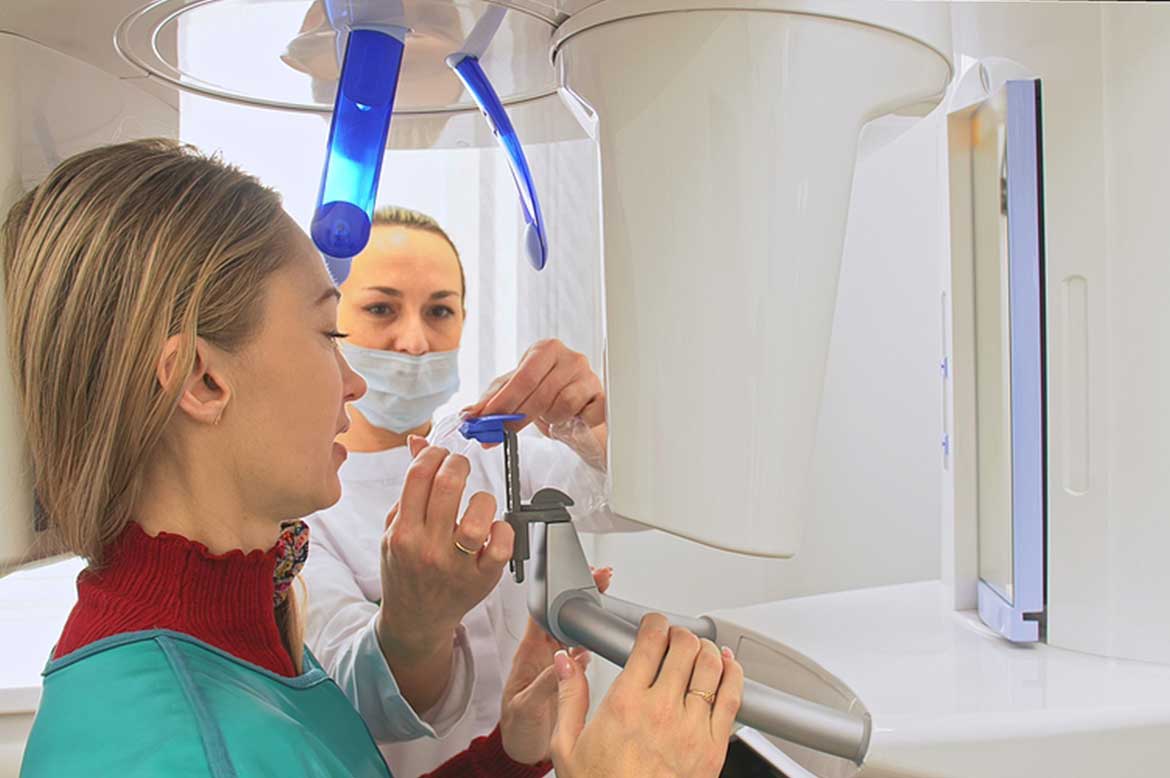
Digital X-rays allow for a two-dimensional view of a three-dimensional object, which is sufficient for many diagnostic purposes. Cone Beam CT technology captures three-dimensional pictures of the teeth and surrounding bone. Your dentist may recommend a CBCT for various applications including dental implant planning, evaluation of the jaws and face, endodontic (root canal) diagnosis and treatment, or diagnosis of dental trauma. This new imaging technology is easy, comfortable, and provides details that help us diagnose and treatment plan optimally.
Digital Intraoral Scanner
Our scanners allow us to take a digital impression, showing a model of your teeth and soft tissues. This means we can generally avoid using the goopy, messy material that has traditionally been used to take impressions. Depending on your needs, we may use these models to study your case, design replacement teeth, plan orthodontic movements, make nightguards and surgical guides for implant placement, and communicate with the lab. The scanners make the process more comfortable and more accurate.
Extraoral and Intraoral Photos
Photography helps us visualize and communicate with you, with our lab, and in diagnosing and treatment planning as we prepare your case. Photography allows us to see cracks or other fragile areas in the teeth or existing restorations, failing margins of fillings, the color, shape, and balance of your teeth and smile, and to document if there is a concerning soft tissue pathology or abnormality. We can compare photos over time if we are monitoring an area.
Would you like to learn more about Imaging?
